Cruet的問題,透過圖書和論文來找解法和答案更準確安心。 我們找到下列問答集和整理懶人包
Cruet的問題,我們搜遍了碩博士論文和台灣出版的書籍,推薦Perez-cruet, Mick J., M.d. (EDT)/ Fessler, Richard G., M.D., Ph.寫的 An Anatomic Approach to Minimally Invasive Spine Surgery 和Herman, Michael的 Wedgwood Jasper Ware: A Shape Book and Collector’s Guide都 可以從中找到所需的評價。
另外網站Salt/pepper cruet Buffo | E.M. Group International - LUSINI也說明:Classic cruet with salt and pepper shaker and jar for toothpicks. Frame and lid made of chrome stainless steel 18/0, rustproof. Dishwasher safe.
這兩本書分別來自 和所出版 。
臺北醫學大學 牙醫學系碩士班 王慶順所指導 VALLEJO FREIRE, LISSETTE SAMANTHA的 A 3D Peptide-based Hydrogel with Anti-inflammatory Effects to Treat Degenerative Disc Disease (2021),提出Cruet關鍵因素是什麼,來自於intervertebral disc、degeneration、functional peptide、self-assembly、hydrogel、compatibility。
而第二篇論文臺北醫學大學 細胞治療與再生醫學國際博士學位學程 蔡 伊琳所指導 Ageng Brahmadhi的 Application of Liquid Chromatography-Mass Spectrometry Methods to Therapeutic Drug Monitoring and Exosomal Proteomics Study (2020),提出因為有 Dried plasma spot、Fluoroquinolones、Microwave-assisted extraction、Therapeutic drug monitoring、Exosome、Proteomic、Placental mesenchymal stem cell、Mass spectrometry的重點而找出了 Cruet的解答。
最後網站The 1000 Islands Cruet則補充:Welcome to the 1000 Islands Cruet. CURRENT STORE HOURS: We are now closed for the season. Re-opening mid-March 2022. Phone: (315) 767-1064.
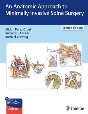
An Anatomic Approach to Minimally Invasive Spine Surgery
為了解決Cruet 的問題,作者Perez-cruet, Mick J., M.d. (EDT)/ Fessler, Richard G., M.D., Ph. 這樣論述:
Learn state-of-the-art MIS techniques from master spine surgeons Significant advances have been made in minimally invasive spine (MIS) surgery approaches, techniques, and innovative technologies. By preserving normal anatomic integrity during spine surgery, MIS approaches enable spine surgeons to
achieve improved patient outcomes, including faster return to normal active lifestyles and reduced revision rates. Exposing only the small portion of the spine responsible for symptoms via small ports or channels, requires a deep understanding of spinal anatomy and spinal pathophysiology. Building
on the widely acclaimed first edition, An Anatomic Approach to Minimally Invasive Spine Surgery, Second Edition, provides an expanded foundation of knowledge to master minimally invasive spine surgery.World-renowned spine neurosurgeons Mick Perez-Cruet, Richard Fessler, Michael Wang, and a cadre of
highly regarded spine surgery experts provide masterful tutorials on an impressive array of cutting-edge technologies. Organized by seven sections and 51 chapters, the book presents a diverse spectrum of current safe and efficacious MIS procedures and future innovations. Nonsurgical approaches inclu
de injection-based spine procedures and stereotactic radiosurgery. Surgical technique chapters discuss MIS anterior, posterior, and lateral approaches to the cervical, thoracic, and lumbar spine, with procedures such as endoscopic microdiscectomy, vertebroplasty and kyphoplasty, percutaneous instrum
entation, and robotic spine surgery.Key FeaturesStep-by-step illustrations, including more than 400 depictions by master surgical and anatomic illustrator Anthony Pazos portray the surgeon's-eye-view of anatomy, intraoperative images, and surgical instruments, thereby aiding in the understanding of
anatomy and procedures20 online videos feature real-time operative fluoroscopy, pertinent anatomy, operative set-up, and common cervical, thoracic, and lumbar approachesDiscussion of novel MIS techniques reflected in 16 new or expanded chapters, including Robotic Assisted Thoracic Spine Surgery and
Stem-Cell Based Intervertebral Disc RestorationThere is truly no better clinical reward for spine surgeons than giving patients suffering from debilitating spinal disorders their life back. This quintessential MIS surgery resource will help surgeons and clinicians accomplish that goal.This book incl
udes complimentary access to a digital copy on https: //medone.thieme.com.
Cruet進入發燒排行的影片
A 3D Peptide-based Hydrogel with Anti-inflammatory Effects to Treat Degenerative Disc Disease
為了解決Cruet 的問題,作者VALLEJO FREIRE, LISSETTE SAMANTHA 這樣論述:
Cell-based therapies for mitigating inflammation-induced damage of intervertebral discs hold promise for the treatment of degenerative disc disease, a condition that is strongly linked to lower back pain. Inspired by marine sandcastle worm’s adhesive protein, we de novo synthesize a self-assembling
peptide hydrogel (PC4) as a three-dimensional (3D) scaffold for nucleus pulposus (NP) tissue engineering. After 3D culture of NP cells in the PC4 hydrogel, we found significant inhibition of proinflammatory responses under LPS stimulation (TNFa, IL-1b and IL-6) as compared to 2D condition. In contr
ast, upregulation of anti-inflammatory genes (IL-10 and Arg-1) and NP signature genes (KRT19 and ACAN) were found in NP cells cultured in 3D PC4 hydrogel as compared to 2D condition, confirming that the system could restore the NP phenotype following de-differentiation during monolayer culture. Cell
viability was high throughout all culture conditions, indicating PC4 hydrogel is biocompatible. Results supported the hypothesis that the 3D PC4 hydrogel could be used as a cell delivery system and scaffold for the treatment of degenerative disc disease.
Wedgwood Jasper Ware: A Shape Book and Collector’s Guide
A PHP Error was encountered
Severity: Warning
Message: file_put_contents(/var/www/html/prints/public/images/books_new/F01/023/37/F010237881.jpg): failed to open stream: No such file or directory
Filename: helpers/global_helper.php
Line Number: 140
Backtrace:
File: /var/www/html/prints/application/helpers/global_helper.php
Line: 140
Function: file_put_contents
File: /var/www/html/prints/application/views/article_v2.php
Line: 248
Function: coverWebp_online
File: /var/www/html/prints/application/controllers/Pages.php
Line: 662
Function: view
File: /var/www/html/prints/public/index.php
Line: 319
Function: require_once
A PHP Error was encountered
Severity: Warning
Message: getimagesize(/var/www/html/prints/public/images/books_new/F01/023/37/F010237881.jpg): failed to open stream: No such file or directory
Filename: helpers/global_helper.php
Line Number: 62
Backtrace:
File: /var/www/html/prints/application/helpers/global_helper.php
Line: 62
Function: getimagesize
File: /var/www/html/prints/application/helpers/global_helper.php
Line: 142
Function: coverWebp
File: /var/www/html/prints/application/views/article_v2.php
Line: 248
Function: coverWebp_online
File: /var/www/html/prints/application/controllers/Pages.php
Line: 662
Function: view
File: /var/www/html/prints/public/index.php
Line: 319
Function: require_once
A PHP Error was encountered
Severity: Notice
Message: Trying to access array offset on value of type bool
Filename: helpers/global_helper.php
Line Number: 64
Backtrace:
File: /var/www/html/prints/application/helpers/global_helper.php
Line: 64
Function: _error_handler
File: /var/www/html/prints/application/helpers/global_helper.php
Line: 142
Function: coverWebp
File: /var/www/html/prints/application/views/article_v2.php
Line: 248
Function: coverWebp_online
File: /var/www/html/prints/application/controllers/Pages.php
Line: 662
Function: view
File: /var/www/html/prints/public/index.php
Line: 319
Function: require_once
A PHP Error was encountered
Severity: Notice
Message: Trying to access array offset on value of type bool
Filename: helpers/global_helper.php
Line Number: 66
Backtrace:
File: /var/www/html/prints/application/helpers/global_helper.php
Line: 66
Function: _error_handler
File: /var/www/html/prints/application/helpers/global_helper.php
Line: 142
Function: coverWebp
File: /var/www/html/prints/application/views/article_v2.php
Line: 248
Function: coverWebp_online
File: /var/www/html/prints/application/controllers/Pages.php
Line: 662
Function: view
File: /var/www/html/prints/public/index.php
Line: 319
Function: require_once
A PHP Error was encountered
Severity: Notice
Message: Trying to access array offset on value of type bool
Filename: helpers/global_helper.php
Line Number: 68
Backtrace:
File: /var/www/html/prints/application/helpers/global_helper.php
Line: 68
Function: _error_handler
File: /var/www/html/prints/application/helpers/global_helper.php
Line: 142
Function: coverWebp
File: /var/www/html/prints/application/views/article_v2.php
Line: 248
Function: coverWebp_online
File: /var/www/html/prints/application/controllers/Pages.php
Line: 662
Function: view
File: /var/www/html/prints/public/index.php
Line: 319
Function: require_once

為了解決Cruet 的問題,作者Herman, Michael 這樣論述:
A lavishly illustrated collector's volume, this book is a wonderful introduction to the historic and ever-popular line of Wedgwood ceramics called Jasper Ware. The bas-reliefs on matte porcelain grounds make these products instantly recognizable. Featuring fine pieces from private and museum collect
ions, it has been written especially for novice and moderately advanced collectors and concentrates on pieces produced mostly from the mid-nineteenth to the early twentieth centuries. Included are chapters on Wedgwood Jasper history, colors, and marks as well as supplements about Wedgwood Jasper jew
elry and the classical mythology used for the bas-relief figures. A significant portion of the book illustrates many of the hundreds of shapes that Wedgwood produced, including biscuit barrels, bud vases, candlesticks, cruet sets, bowls, inkwells, and jardini res, to name only a few. Over 500 vivid
photographs illustrate these shapes, and detailed information as well as current values are included in each caption. This is an important book about a time period in Wedgwood Jasper history that has not been researched before. It will be a welcomed addition to the library of all Wedgwood Jasper ent
husiasts. Michael Herman is a retired teacher and librarian. Wedgwood collecting is a hobby that he and his wife share with equal ardor and both are members of the Wedgwood Societies of New York and Washington D.C. as well as the Wedgwood Friends of CT/NY.
Application of Liquid Chromatography-Mass Spectrometry Methods to Therapeutic Drug Monitoring and Exosomal Proteomics Study
為了解決Cruet 的問題,作者Ageng Brahmadhi 這樣論述:
TABLE OF CONTENTCOVER PAGETHESIS CERTIFICATIONACKNOWLEDGEMENT ITABLE OF CONTENT IIILIST OF TABLES VILIST OF FIGURES VIILIST OF ABBREVIATIONS VIIIOVERVIEW OF PhD DISSERTATION XPART I. 1Liquid chromatography-mass spectrometry 1Chapter 1. Introduction of Liquid chromatography-mass spectrometry
21.1. Liquid Chromatography 21.2. Mass Spectrometry 41.3. Ion Source 41.3.1. Atmospheric Pressure Chemical Ionization (APCI) 51.3.2. Electrospray ionization (ESI) 61.4. Mass analyser 61.4.1. Quadrupole mass spectrometer 71.4.2. Time of flight mass spectrometers 81.4.3. Quadrupole ion-
trap spectrometers 81.4.4. Fourier-transform ion cyclotron mass spectrometers 91.4.5. Orbitrap mass analyser 91.5. Liquid chromatography–mass spectrometry application 10PART II 11Application of Liquid Chromatography-Mass Spectrometry Methods to Therapeutic Drug Monitoring 11Abstract 12Chapt
er 2: Introduction of therapeutic drug monitoring for anti-TB treatment 14Chapter 3: Literature review of therapeutic drug monitoring for anti-TB treatment 163.1. Tuberculosis burden 163.2. Fluoroquinolones 173.3. Therapeutic drug monitoring 183.4. Dried plasma spots 193.5. Microwave-assisted
extraction (MAE) 19Chapter 4: Material and Methods of therapeutic drug monitoring for anti-TB treatment 214.1. Reagents and chemicals 214.2. DPS, plasma sample preparation and MAE 214.3. Method validation dried plasma spot 234.3.1. Selectivity 234.3.2. Calibration curves and quality contr
ol samples 234.3.3. Accuracy and precision 234.3.4. Extraction recovery and matrix effect 234.3.5. Stability and carryover 244.3.6. The dilution integrity 244.4. Plasma sample type method validation 244.5. Clinical application 25Chapter 5: Results and Discussions of therapeutic drug mon
itoring for anti-TB treatment 265.1 Sample extraction 265.2 UHPLC-MS/MS 295.3 Dried plasma spot method validation 305.3.1 Selectivity 305.3.2 Calibration curves, accuracy and precision 305.3.3 Recovery, matrix effect, stability and carryover 325.3.4 The dilution Integrity 335.4 Plasm
a sample type method validation 335.4.1 Selectivity 345.4.2 Calibration curve and quality control 346. Clinical application 36Chapter 6. Conclusion 40PART III 41Application of Liquid Chromatography-Mass Spectrometry Methods to Exosomal Proteomic Study 41Abstract 42Chapter 7: Introduction
of exosomal proteomic study 43Chapter 8: Literature review of exosomal proteomic study 448.1. Mesenchymal stem cells 448.2. Mesenchymal stem cells and immune system regulation 458.3. Placental Mesenchymal Stem Cells (pcMSCs) 478.4. Exosome 478.4.1. Exosome definition and size coverage 478.4.
2. Exosome biogenesis 488.5. Exosome isolation and characterization 49Chapter 9: Material and methods of exosomal proteomic study 519.1. Material and reagents 519.2. Initial centrifugation of conditioned medium 519.3. Centrifugal concentration of conditioned medium 519.4. Exosome isolation by
size exclusion chromatography 529.5. ExoQuick-TC™ exosome precipitation solution isolation 529.6. BCA Protein Assay 529.7. Tunable resistive pulse sensing 539.8. Electron microscopy 539.9. Western Blot Analysis 539.10. SDS-PAGE, In-Gel Digestion, Mass spectrometry (MS) 549.11. Bioinformatics
Analysis 54Chapter 10: Results and discussions of exosomal proteomic study 5510.1. Exosome isolation 5510.2. Exosome characterization 5610.3. Proteomic of the pcMSC exosome 5810.4. Exosome contents and skin wound healing 73Chapter 11: Conclusion 78References 79LIST OF TABLESTable 1. Multipl
e reaction monitoring parameters of three fluoroquinolones, and the internal standard (moxifloxacin hydrochloride-13CD3). 22Table 2. Calibration curves of the fluoroquinolone. 31Table 3. The LLOQ and QC samples accuracy and precision of levofloxacin, ciprofloxacin, moxifloxacin. 32Table 4. Extrac
tion recovery and matrix effect of QC samples for levofloxacin, ciprofloxacin, moxifloxacin. 32Table 5. Auto-sampler (4℃) and four-day (-80℃ and room temperature) stability of levofloxacin, ciprofloxacin, moxifloxacin. 33Table 6. Calibration curves of levofloxacin, ciprofloxacin, moxifloxacin for
plasma samples. 34Table 7.Accuracy and precision of quality control plasma samples for the three target analytes. 35Table 8.Comparison of the accuracies and precisions of quality control samples from DPS and plasma preparations. 36Table 9. Drug concentrations of clinical samples in dried plasma s
pot (DPS) and plasma. 37Table 10. Exosome size coverage range 48Table 11. BCA standard preparation 53Table 12. List of identified protein in exosome sample, exocarta, and vesiclepedia. 60Table 13. The 25 most relevant pathways and involved proteins 65Table 14. DAVID functional annotation of ide
ntified protein 68Table 15. DAVID Functional clustering 71Table 16. MMP protein family in skin wound healing process 75Table 17. Dynamic changes of MMP types and scars conditions (148) 76LIST OF FIGURESFigure 1. Diagrammatic of separation (2). 3Figure 2. General layout of mass spectrometers (5)
. 4Figure 3. Schematic diagram of atmospheric pressure chemical ionization source (6). 5Figure 4. Schematic diagram of Electrospray ionization source (6). 6Figure 5. Schematic diagram of quadrupole system (9) 7Figure 6. Schematic diagram of MALDI field-free drift region (6) 8Figure 7. Schematic
diagram of quadrupole ion-trap spectrometers (11) 9Figure 8. Anatomy of orbitrap mass analyser. 10Figure 9. Fluoroquinolones molecular structures. 17Figure 10. Microwave-assisted extraction (400 W) peak area percentages of the three analytes at different extraction times.. 27Figure 11. Microwav
e-assisted extraction optimization parameters. 28Figure 12. Chromatogram of three analytes extracted with MAE 400 W for 40 seconds in different solvent (90% methanol, 90% acetonitrile and 90% isopropanol). 29Figure 13. The overlaid chromatograms of plasma selectivity 30Figure 14. The selectivity
of plasma preparation.. 34Figure 15. Deming regression and Bland-Altman plot of drugs concentration in DPS and plasma. 39Figure 16. Sensor and switcher model of MSC (94) 45Figure 17. MSC balancing macrophage polarization into anti and pro inflammatory phenotypes (92) 46Figure 18. Exosome Biogene
sis 49Figure 19. Overview of exosomal study design 51Figure 20. Exosome isolation workflow. 55Figure 21. Exosome characterization workflow.. 56Figure 22. Exosome characterization 56Figure 23. Concentration and particle diameter of isolated exosomes. 57Figure 24. TEM visualization of the isolat
ed exosome.. 58Figure 25. A. SDS-PAGE of exosome derived protein. B. chromatogram of the selected fraction 59Figure 26. Venn diagram of identified proteins. In contrast to the two databases 60Figure 27. Cellular component for exosome samples 63Figure 28. Molecular functions for exosome samples
64Figure 29. Biological process for exosome samples 65Figure 30. Time scale of the four phases of wound healing process (141) 74